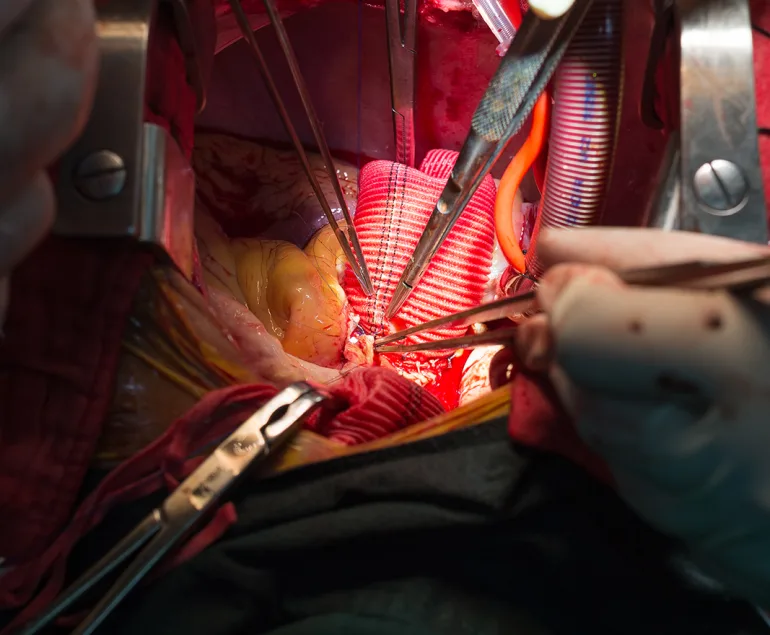
How is Aortic Aneurysm Diagnosed?
Aortic aneurysms are usually asymptomatic, especially if they are small, and may even be asymptomatic if they are very advanced. As the aortic aneurysm enlarges, it can cause pain. Depending on the location of the aneurysm, the pain may occur in the anterior chest wall, back, anterior abdominal wall or lower back. The pain is usually vague, very severe pain only occurs when the aneurysm ruptures. In some cases, aortic aneurysm disease can progress suddenly and rapidly without any symptoms. In such cases, patients without symptoms may not realise that they have an aortic aneurysm until they see a doctor. In asymptomatic patients, aortic aneurysm disease is usually discovered by chance during a routine check-up or scan.
Especially hypertensive patients, people over a certain age, people with aortic aneurysm disease in their family are at risk. People in these risk groups can consult their doctor and find out whether they have aortic aneurysm disease thanks to the common methods used.
Common methods used to diagnose aortic aneurysm can be listed as follows:
- Physical examination: Aneurysm especially in the abdominal region can be diagnosed by physical examination performed by the doctor. However, physical examination alone is not sufficient for diagnosis, it should be supported by imaging tests.
- Imaging Tests:
- Ultrasound Ultrasound can be used to visualise the condition of the aorta and other vessels and to determine whether there is an enlargement in the diameter of the aorta. It can be used to assess whether there is any damage or weakening of the aortic wall. Ultrasound is one of the common methods used to detect aortic aneurysms. Ultrasound is the first diagnostic method used especially in abdominal aortic aneurysms.
- Echocardiography: It is the first test used especially in the diagnosis of ascending aortic aneurysms. A good echocardiographic examination can also provide information about aneurysms of the arch aorta and descending aorta. Echocardiography is also the first-line imaging method in the follow-up of ascending aortic aneurysms.
- Computed Tomography (CT) Scan: Computed tomography scan provides more detailed images of the aortic vessel and diagnosis is made. CT scan not only provides information about the aneurysm, but also gives us valuable information about the surrounding tissues. Computed tomography is used to determine the size of the aneurysm, the thickness of the aortic wall, the current condition of the tissues surrounding the aortic aneurysm, and whether there is bleeding around the aneurysm. Thus, possible complications that may occur in other organs are tried to be determined and the treatment process is initiated after diagnosis.
- Magnetic Resonance Imaging (MRI): Magnetic resonance imaging method provides high quality images and allows inferences to be made about the current condition of the aorta. It is used to better determine the severity, structure and extent of aneurysm enlargement. With the help of MR, possible complications are detected and the treatment process is initiated.
- Aortic Angiography Aortic angiography provides a detailed visualisation of the current condition of the aorta and the severity of the aneurysm. Aortic angiography is an invasive procedure. Therefore, there are risks such as vascular injury, infection, allergic reactions due to contrast material.
Even if you do not have symptoms of aortic aneurysm, you can consult a Cardiovascular Surgery specialist to have the necessary tests performed and start the treatment process early according to the results.