How is the Ozaki Surgery Performed?
The Ozaki Procedure, first performed by Japanese scientist Dr. Shigeyuki Ozaki in 2007, is an aortic valve replacement method that has been performed in Turkey since 2018. Considering the advancements in cardiac surgery and hospital infrastructure in Turkey, it is reasonable to expect an increase in the use of modern and innovative treatment methods like the Ozaki procedure.
The Ozaki surgery is a surgical procedure used to create new aortic valves in patients with aortic valve stenosis and aortic valve insufficiency. During the surgery, either the patient’s own heart membrane is removed or a specially produced pericardium from a cow’s heart membrane is used to create new aortic valves. In both cases, the patient’s aortic valve is created during surgery and is completely customized for the patient, containing no foreign materials except for the suture material. For these procedures, the patient is connected to a heart-lung machine, the heart is stopped, the aortic valves are removed, the aortic annulus is cleaned, and the prepared new valves are sutured into place.
During the surgery, a heart-lung machine may be used outside the heart to temporarily take over its function and regulate blood circulation in the body. To begin the procedure, the heart is stopped using a special solution (cardioplegic solution). This solution temporarily stops the heart, allowing the surgeon to perform the surgery smoothly.
The surgeon frees and removes the patient’s own heart membrane to create a new heart valve for the patient. The patient’s damaged valves are removed, and measurements are taken for the new valves. Valves suitable for the patient are cut from the patient’s own heart membrane or from a prepared bovine heart membrane.
After the new Ozaki valves are implanted into the patient, the incisions are closed, and the patient’s heart is restarted and disconnected from the heart-lung machine. The newly implanted valves are evaluated using transesophageal echocardiography. Once no issues are confirmed, the patient’s incisions are closed, and the surgery is completed, with the patient being transferred to intensive care for monitoring.
The Ozaki procedure is performed step by step as follows:
Preparation Process
The patient must undergo a detailed evaluation before surgery. This evaluation usually includes blood tests, imaging studies, and other medical tests. In addition, the patient receives a series of instructions before surgery, such as fasting and avoiding certain medications.
General Anesthesia
On the day of surgery, the patient arrives at the operating room and is placed under general anesthesia. This causes the patient to lose consciousness and not feel anything during the surgery.
Incision and Access to the Aortic Valve
Access to the heart is gained through the rib cage or between the armpits. A piece of the heart membrane of the appropriate size is removed and placed on the operating table for the procedure. It is then immersed in a special solution.
Cannulas are placed to connect the patient to the heart-lung machine, and the patient is connected to the heart-lung machine and placed on cardiopulmonary bypass.
Preparation of the Aortic Valve
The heart is stopped, and an aortotomy is performed to reach the aortic valve leaflets. The aortic valve leaflets are resected, and measurements are taken for the new valve leaflets.
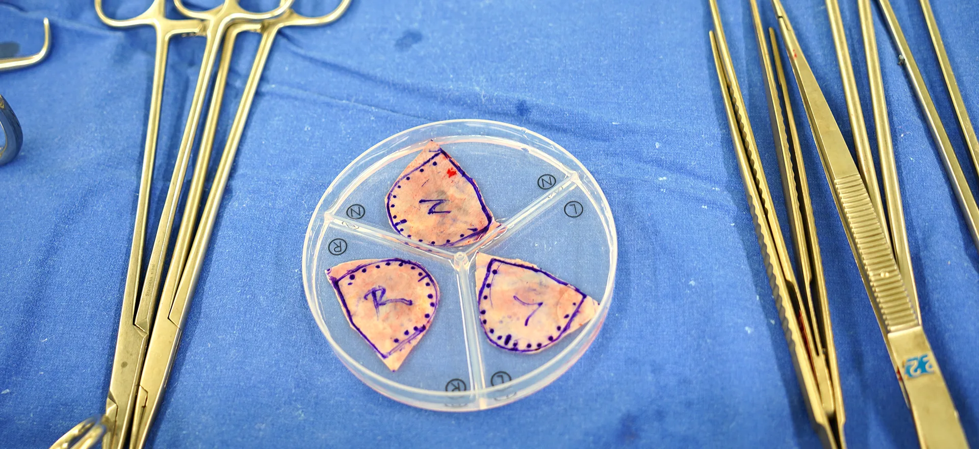
Creation and Placement of the New Valve
The surgeon completes the preparation of the heart membrane on the other table and makes it ready for use. New valves are created according to the aortic valve measurements.
The valves are implanted into the aortic annulus using a special suturing technique, each in its own area. No other material is used besides sutures when attaching the valves.
Testing the Valve
After the Ozaki valves are sutured into place, the aortic incision is closed, and the patient is warmed and disconnected from the heart-lung machine. The new Ozaki valves are checked using the transesophageal echocardiography placed at the beginning of the surgery. Once no problems are confirmed, the patient’s cannulas are removed and the closure phase begins.
Closure of Incisions
After the cannulas are removed, bleeding is controlled, drains are placed, and the incisions are closed according to procedure, completing the surgery.
Postoperative Care
The patient is admitted to the intensive care unit and closely monitored after surgery. The patient usually stays in the intensive care unit for one day and is transferred to the ward the next day.
Rehabilitation and Recovery
The patient is admitted to the ward with a drain and some cannulas. The patient is mobilized and given breathing exercises while in the ward. On the second day in the ward, the drains and catheters are removed, and the patient is able to move around and take care of their needs independently. After the fourth day in the ward, the patient’s parameters are evaluated, and they are discharged.
Follow-up and Monitoring
Routine check-ups are performed one week and one month after discharge, followed by six-monthly check-ups.
This process may vary depending on the patient’s underlying conditions, the complexity of the surgery, and personal factors. It is important for the patient to rest and follow the doctor’s recommendations. Additionally, it is important not to miss doctor’s appointments, monitor the process, and ensure that any necessary additional measures are taken and medications are adjusted as needed.